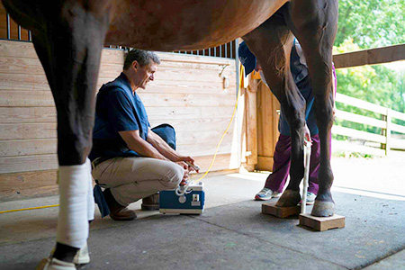
We regularly invest in up-to-date equipment and training, to offer the most current diagnostic techniques available. Advances diagnostics and treatment are made every year and at Running ‘S’ Equine, we stay current by upgrading our equipment, attending continuing education seminars and advancing our skill sets, for the benefit of your horse.
Digital Radiography
Digital Radiography provides immediate acquisition of diagnostic images of the highest quality. In the hospital, a powerful, overhead-suspended high frequency radiograph machine allows us to obtain diagnostic quality images of larger body parts, such as the head, pelvis, chest and abdomen.
Digital Ultrasonography
Our Digital Ultrasonography equipment resolves structures as small as one millimeter! Color-flow Doppler ultrasonography is available for advanced examination of the heart and other vascular (blood vessel) structures.


Video Endoscopy and Gastroscopy
We offer digital video endoscopy and video gastroscopy of the upper airways and the esophagus, stomach and proximal small intestine. These procedures can be performed in the hospital or in the field at the horse’s location, in most cases. Endoscopic biopsies are obtained, and foreign bodies grasped and removed with special endoscopic instruments. Our endoscopes are also used to visualize the lower intestinal tract and the urethra and bladder. They are sometimes even used to assist with laparoscopy!
Digital Venography and Arthrography
Digital Venography is a radiographic technique to image the blood supply of the horse’s foot. Radiographic contrast material is injected into the local blood vessels and a series of radiographs are obtained. This technique offers a rapid, cost-effective method of evaluating the blood flow to the foot in cases of laminitis or in the diagnosis of other conditions like infections or keratomas. The added expense of Magnetic Resonance (MRI) is often avoided with this technique.


Arthrography is used to view cartilage defects in joints. Standing (low field) MRI cannot image joint cartilage, so cartilage lesions and defects go undetected. Arthrography, is an imaging procedure in which radiographic contrast material is injected into the joint, can be used to confirm cartilage lesions and other joint and bone-related pathology.


Tonometry
Rapid and accurate measurement of the pressure inside the eye (Intraocular Pressure IOP) is made with our computerized TONOPEN. Glaucoma can be diagnosed early and treatment initiated, avoiding or slowing the development of complications and additional eye damage.
Arthroscopy, Laparoscopy, Tenoscopy and Bursoscopy (Tendon Sheathes and Bursas)
Sometimes, the best way to diagnose what is going on inside a joint, the abdomen, a tendon sheath or bursa is simply to take a look! Minimally invasive techniqes, using tiny incisions, a small telescope with a digital video image projected on a video monitor can offer what other imaging techniques cannot; a direct look. Small instruments, specifically-designed for this type of procedure can be used to take biopsies, remove foreign bodies or correct abnormalities at the time of the exploratory procedure.
Exploratory Surgery
Open, exploratory surgery is also often necessary. The abdomen and sinuses are the most common sites for this type of surgery.
Referral Services
Other diagnostic services such as Magnetic Resonance Imaging (MRI), nuclear scintigraphy and Computed Tomography (CT) are available on a referral basis, with expert consultation by Board Certified Radiologists and other Specialists.
Imaging Consultations
Looking for your next horse? Need a second opinion? Running ‘S’ Equine Veterinary Services offers complete imaging consultations. Dr. Staller views radiographs, ultrasonography images, endoscopy images, MRI’s Bone Scans, Venograms, and Mylograms.
